About the clinic
Lower back pain is a common but very often underdiagnosed presentation in small animal practice. Spotting that something is wrong is the easy part, but working out where the problem originates can be much harder. A dog that is reluctant to jump, slow to rise or vaguely stiff behind can be examined carefully and still leave differentials spanning spinal disease (lumbosacral disease, IVDD, vertebral malformation or instability, neoplasia), hip pathology (osteoarthritis, dysplasia) and muscular or soft tissue pain (iliopsoas injury, degenerative or inflammatory muscle disease).
These conditions overlap and mimic one another, and plain radiographs will miss the majority of them. Plain films may confirm hip disease, for example, while the neurological disease driving the signs goes unseen.
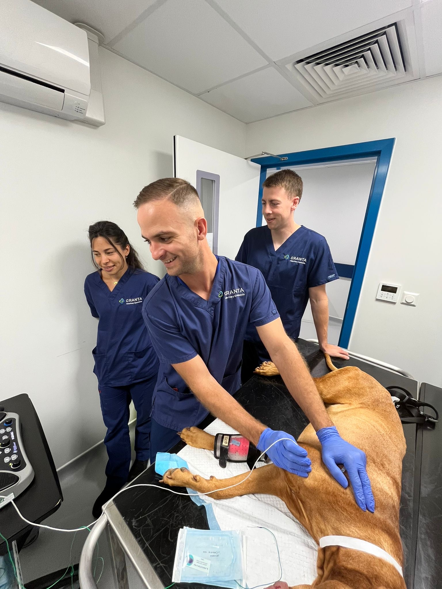
Our lower back pain clinic brings the investigation together in one place. Rather than working through modalities sequentially, we combine MRI, dynamic CT and electrodiagnostic testing in a single visit and under one general anaesthetic, then send every patient home with a tailored treatment plan and a full written report for you.
Who the clinic is for
Dogs where you suspect lower back pain but the cause remains unclear, particularly:
- Reluctance to jump, use stairs or rise from rest
- Vague hindlimb stiffness that is not clearly either orthopaedic or neurological
- Pain localised to the lumbosacral region or hips on examination
- Poor response to empirical pain management protocols
- Differentials spanning spine, hips and muscle with no clear front-runner
- Behavioural change such as new reactivity, restlessness or difficulty settling, alongside subtle hindlimb signs
What the package includes
- MRI, the imaging of choice for the spinal cord, nerve roots, lumbosacral junction and surrounding soft tissues, including muscular lesions such as iliopsoas disease
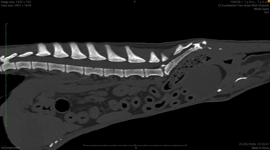
- Dynamic CT, superior bone detail for vertebral anatomy, hip morphology and bony lesions not easily identifiable on plain films, with dynamic acquisition to assess instability or abnormal motion
- Electrodiagnostic testing, EMG and nerve conduction studies to assess nerve and muscle function, support whether imaging findings may be clinically significant and detect some diseases not visible on imaging
After the investigations
Every patient receives a tailored treatment plan, whether that is medical management, physiotherapy, interventional pain management or a surgical discussion, as the findings direct. You receive a full written report with clear follow-up guidance, and we are always happy to discuss a case informally before referral.
When the scans do not tell the whole story
Some of the dogs we see arrive with normal imaging, or with signs that look more like behaviour than pain. Viv Moore, an accredited canine behaviourist and trainer, has brought three of her own dogs to our neurology team with lower back pain. Each reached an answer by a different route, and she has kindly let us share their stories.
Doug, 9½, Labrador
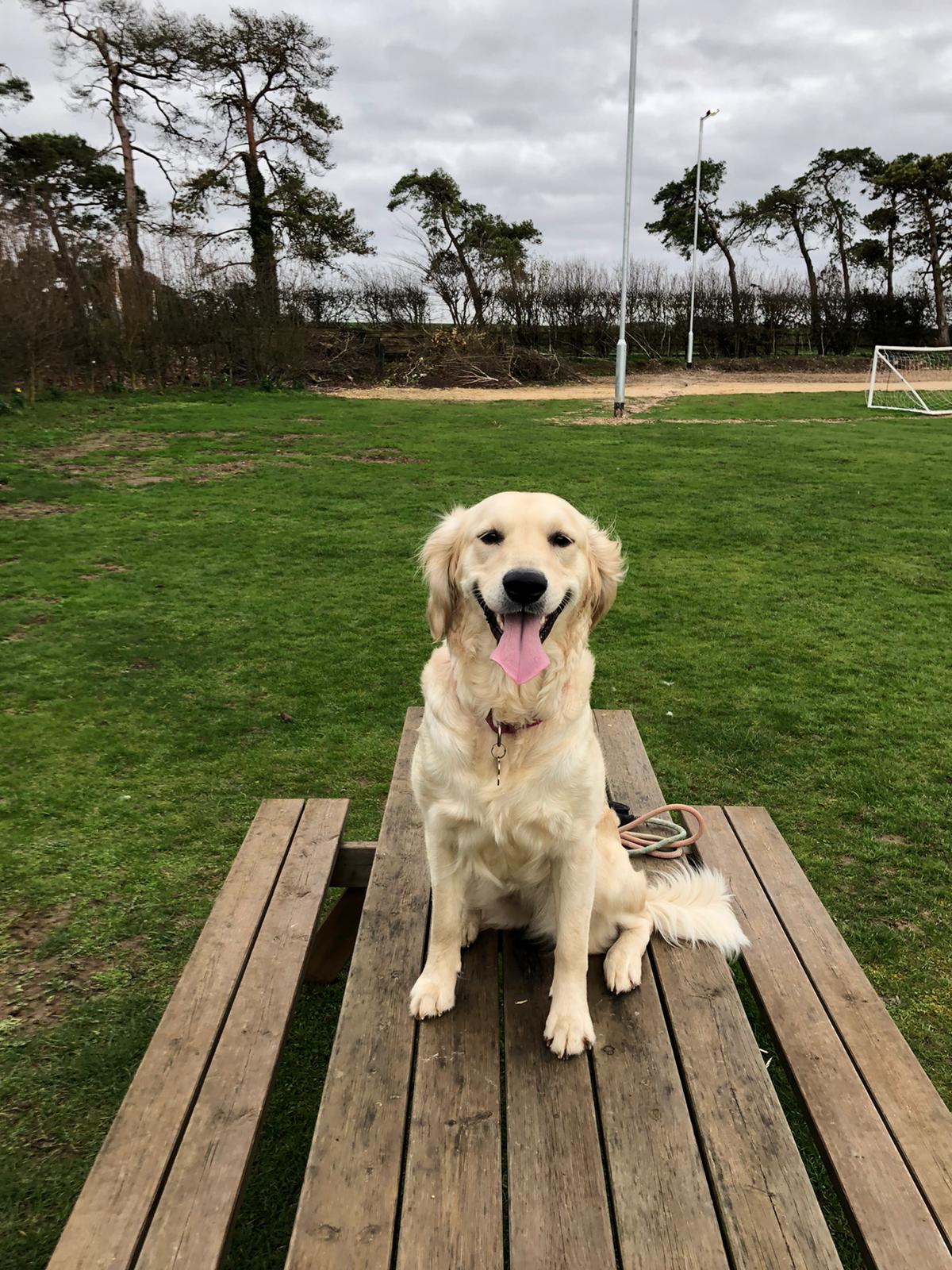
Doug was never lame and never cried out. Over around two years he became more reactive to unfamiliar dogs and people, sensitive to noises at home and distressed when left alone, all of it out of character. The first physical clue was a subtle offloading of weight through his hind limbs, picked up at a behaviour workshop Viv attended with him.
Doug was diagnosed with degenerative lumbosacral stenosis at another referral centre and managed medically for about a year before Viv moved his care to our neurology team. He had not been offered a steroid epidural before, and it proved a turning point. His care is ongoing.
“Following Doug’s first steroid epidural, the change in him was remarkable. It genuinely felt like we had our old Doug back.”
Rosie, 2
Rosie first showed a brief hind limb limp after rest or on walks, lasting only a few seconds. Radiographs of her hips, lumbar spine and stifles showed nothing significant, and she was first seen by our orthopaedic team for a suspected iliopsoas strain. Over the following weeks her behaviour changed. She struggled to settle in the evenings and became much less comfortable around the other dogs at home.
A short video of Rosie holding a hind leg off the ground on a walk led to her transfer to our neurology team. Her dynamic MRI showed no obvious abnormality, but her history, clinical signs and response to treatment raised the possibility of hypermobility in the lumbosacral region. After long conversations about the uncertainty, Rosie had spinal stabilisation surgery, followed by physiotherapy and hydrotherapy. By her second birthday she was running off lead again.
“It felt like she could finally just be a young dog again.”
Bill, 2½
Bill came to Viv as a rescue puppy after a difficult start and struggled to settle from his very first night. As he grew she noticed subtle weight shifting through his left hind limb, an unusual pacing gait, trouble switching off to rest and, when he was younger, episodes of involuntary urination. Anti-inflammatories made little difference, while gabapentin helped noticeably.
Radiographs and a dynamic MRI here showed no obvious structural abnormality, but examination found pain around his lumbosacral region. His first steroid epidural brought a significant improvement. A second helped for a shorter time, and the team are now considering whether tethered cord syndrome could explain the rest. Bill’s story is still unfolding.
“Although Bill’s journey isn’t over yet, we’re incredibly grateful to have a team who continue to listen, investigate and support us while we work towards finding those answers.”
What Viv has learned
“Doug taught me to recognise subtle changes. Rosie taught me that sometimes the whole clinical picture is more important than the scans alone. Bill has taught me that sometimes the answers don’t come quickly.”
Fees and referral
The combined package is £3,400. It covers the consultation, day admission, general anaesthetic, MRI, dynamic CT study, electrodiagnostic testing and a full referral report, offering a significant saving compared with pursuing the same investigations sequentially.
To refer, please use our standard referral form at grantavets.co.uk/referring-vets/book-a-referral. If you would like to talk a case through first, contact hello@grantavets.co.uk FAO Max Foreman.